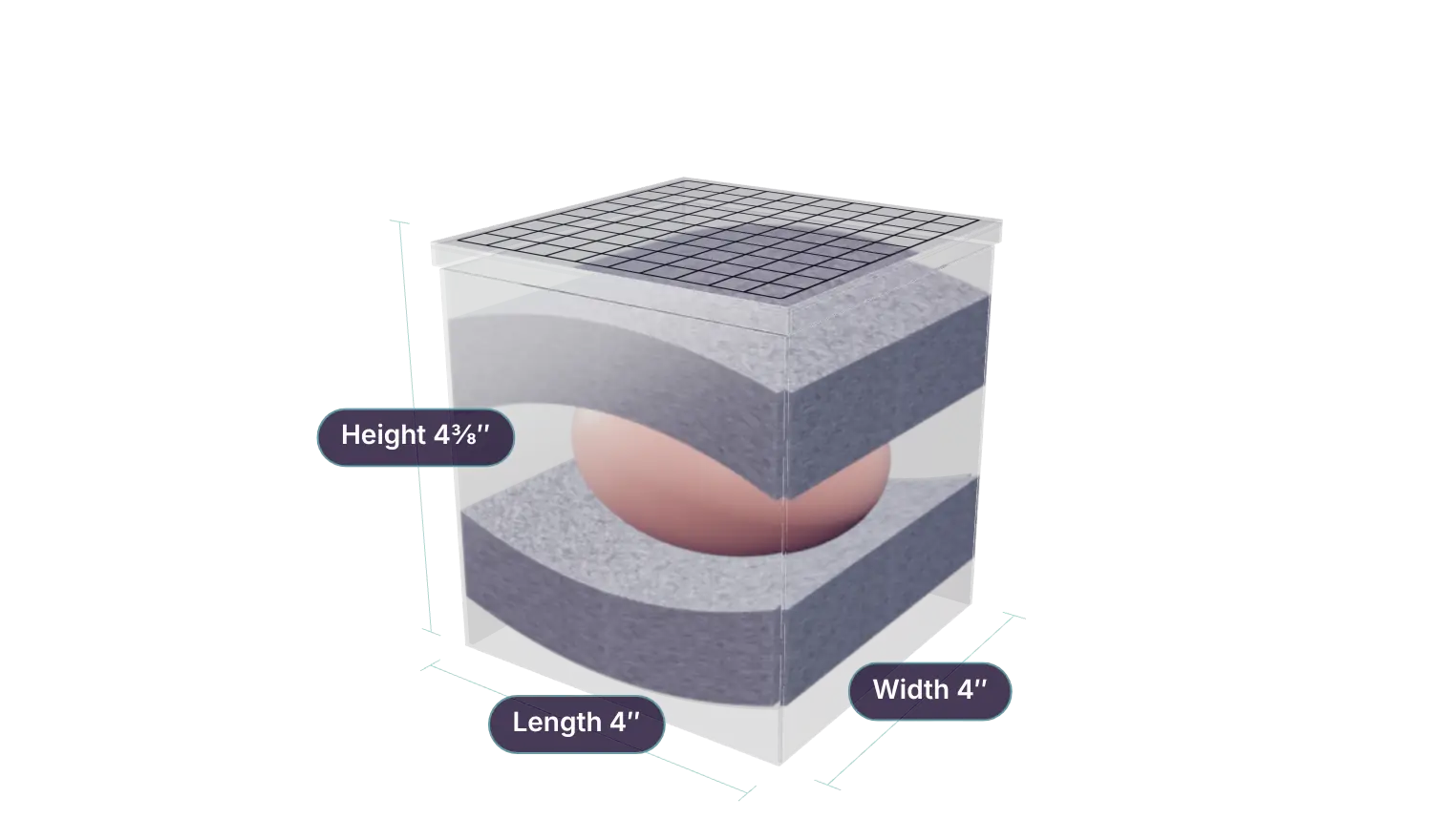
Radio-opaque grid
The grid appears in the upright image and is visible on the lid, helping identify close margins and areas of interest for the pathologist.
Now you can get the orthogonal views you need without risk of specimen damage. The specimen travels from the OR to Imaging to Pathology untouched.
Overall size: 4″ L × 4″ W × 4⅜″ H · lid grid 1 cm squares
Surgeons who remove breast specimens and send them for mammographic interpretation worry that compression or manipulation will either interfere with or damage the margins they have excised. Clinical articles indicate that compression can create false negative margins in some cases (pancake phenomena) or that manipulation of the specimen can create false positive margins by damaging the tissue after excision.
What surgeons need is a mammographic review of their removed specimen that shows it in its “as removed” condition, without damage or compression.
The 2-View provides a method for viewing the specimen in multiple orthogonal views without being compressed or manipulated. Once these views are obtained and reviewed, the radiologist can report with confidence back to the surgeon what the margin conditions are, and reference the grid markings on the container to verify which margins are in question. The specimen continues on to pathology in its original condition, giving the pathologist greater confidence in his margin analysis as well.
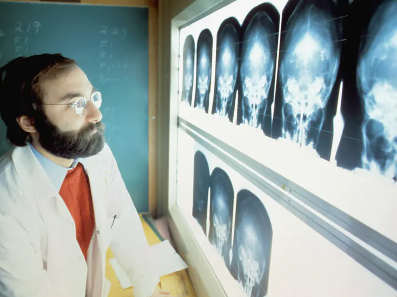
The grid appears in the upright image and is visible on the lid, helping identify close margins and areas of interest for the pathologist.
Take the first view, rotate the device 90 degrees vertically, and take the second. The two views can be compared for a 3-dimensional analysis of the margins.
The specimen continues on to pathology in its original condition, giving the pathologist greater confidence in the margin analysis.
Ask your operating room director or purchasing manager to contact 2-View, LLC, or order a supply of 2-View specimen containers today.